IMAGE

Figure 4

ID
ZDB-IMAGE-210922-8
Source
Figures for Chang et al., 2021
Image
Figure Caption

Figure 4

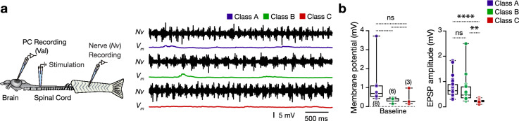
Adult zebrafish valvular Purkinje cell do not discharge during fictive locomotion. (a) Ex-vivo setup of the brain-spinal cord allows simultaneous recordings of Purkinje cells and ipsilateral motor nerves. Sample recordings from each valvular Purkinje cell firing class during locomotion. (b) Analysis of membrane potential depolarization and the amplitude of the recorded EPSPs during locomotion between the different classes of Purkinje cells. Data are presented as box plots showing the median with 25/75 percentile (box and line) and minimum–maximum (whiskers). **P < 0.01; ****P < 0.0001; ns not significant. For detailed statistics, see Supplementary Table S1.

Acknowledgments
This image is the copyrighted work of the attributed author or publisher, and ZFIN has permission only to display this image to its users. Additional permissions should be obtained from the applicable author or publisher of the image. Full text @ Sci. Rep.