IMAGE

Figure 1

ID
ZDB-IMAGE-210916-1
Source
Figures for Chen et al., 2021
Image
Figure Caption

Figure 1

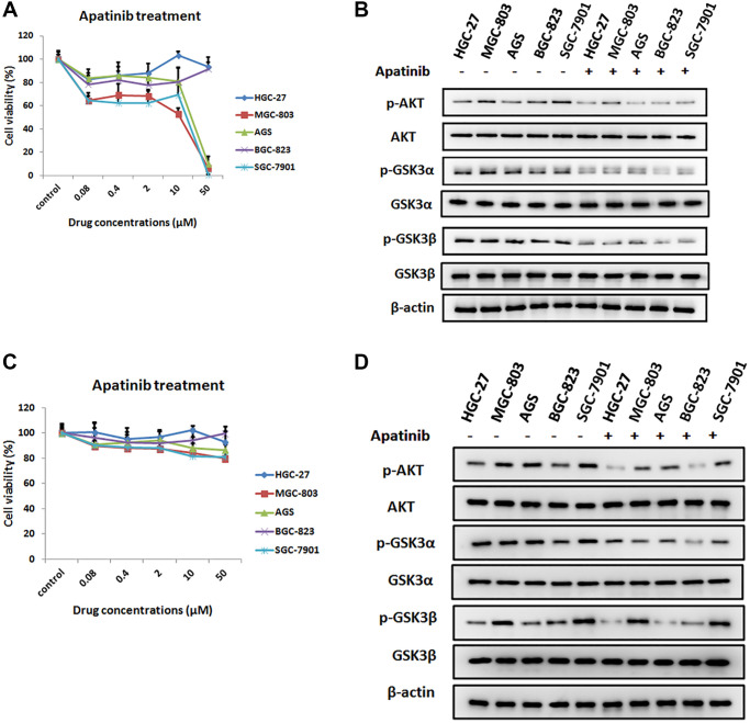
The inhibition effect of apatinib on the cell viability in different GC cell lines. (A) Cell viability of different human gastric cancer cell lines treated with different concentrations of apatinib for 72 h. (B) After treatment with apatinib 2 μM concentration for 72 h, western blot analysis of AKT, phosphorylated AKT (p-AKT), GSK3α, phosphorylated GSK3α (p-GSK3α), GSK3β and phosphorylated GSK3β (p-GSK3β) in GC cell lines. (C) Cell viability of different human gastric cancer cell lines treated with different concentrations of apatinib for 8 h. (D) After treatment with apatinib 2 μM concentration for 8 h, western blot analysis of AKT, phosphorylated AKT (p-AKT), GSK3α, phosphorylated GSK3α (p-GSK3α), GSK3β and phosphorylated GSK3β (p-GSK3β) in GC cell lines.

Acknowledgments
This image is the copyrighted work of the attributed author or publisher, and ZFIN has permission only to display this image to its users. Additional permissions should be obtained from the applicable author or publisher of the image. Full text @ Aging (Albany NY)