
Image
|
Figure Caption
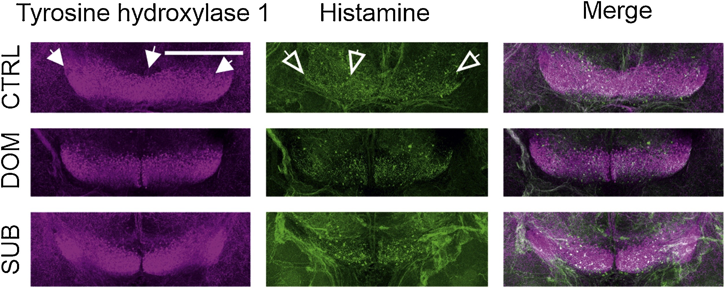
Fig. 9 Immunohistochemistry of histamine and tyrosine hydroxylase positive neurons in the adult male brains of the F1 generation. Throughout the images tyrosine hydroxylase immunoreactivity is depicted in magenta and histamine immunoreactivity is depicted in green. The brains are orientated in a similar manner in all sub-images, with the anterior to the top of the page, the posterior to the bottom of the page and the diencephalon and hypothalamus visualized from the ventral side of the brain. A horizontal view. CTRL = control, DOM = dominant, SUB = subordinate. n = 5/group. Scale bar =250 μm.
Acknowledgments
This image is the copyrighted work of the attributed author or publisher, and
ZFIN has permission only to display this image to its users.
Additional permissions should be obtained from the applicable author or publisher of the image.
Full text @ Behav. Brain Res.