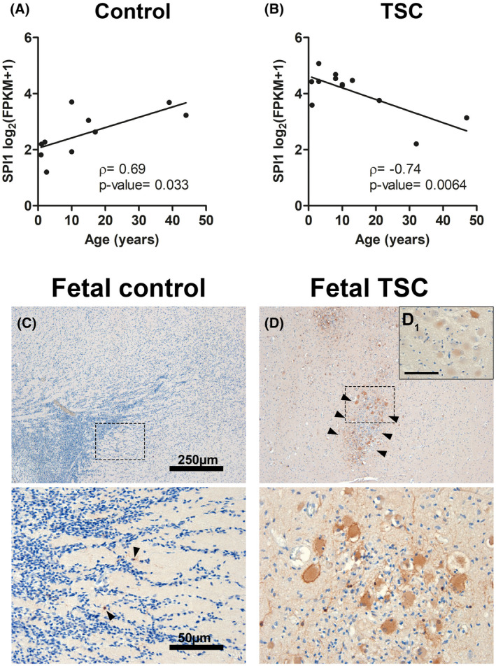
SPI1/PU.1 expression in malformed cells occurs early in TSC development. SPI1 expression displayed a significant positive correlation with age in control samples (A). In contrast, TSC tuber tissue displayed a negative correlation with age samples (Pearson's correlation) (B). Fetal control cortex displayed sparse SPI1/PU.1 expression solely in few microglia (arrowheads in magnified lower panel) (C). Fetal TSC cases revealed strong expression of SPI1/PU.1 in malformed cells that could be found in subcortical clusters (arrowheads, magnified in lower panel) (D). SPI1/PU.1 expression was found to be variable between malformed cells (D1). Sections C and D were counterstained with hematoxylin. Scale bars: 250 µm in upper panel and 50 µm in C (representative for D), 25 µm in D1; A, B Spearman's rank correlation test (displayed with Spearman correlation coefficient ρ and exact p‐value)
Acknowledgments
This image is the copyrighted work of the attributed author or publisher, and
ZFIN has permission only to display this image to its users.
Additional permissions should be obtained from the applicable author or publisher of the image.
Full text @ Brain Pathol
Your Input Welcome
Thank you for submitting comments. Your input has been emailed to ZFIN curators who may contact you if
additional information is required.
Oops. Something went wrong. Please try again later.