
|
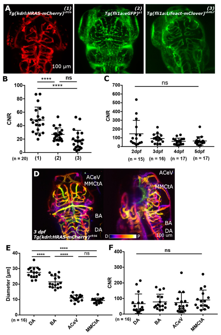
Figure 2
(A) Image quality was assessed by contrast-to-noise ratio (CNR) level measurement in three different transgenic lines, which harbour different reporter constructs (promotor as well as reporter; 1,2,3). (B) CNR levels in these transgenic reporter lines showed a statistically significant difference between (1) and (2) as well as between (1) and (3), with p < 0.0001 (****) for both. Between (2) and (3) no statistically significant difference was found (p 0.0898; ns). (C) CNR levels from 2-to-5 dpf showed no statistically significant difference (p 0.1032; ns). (D) Light sheet fluorescence microscopy (LSFM) allows to visualize vessels from the most dorsal plane (p - proximal image plane; e.g., anterior cerebral vein (ACeV)) to vessels which are a few hundred micrometer inside the embryo (d distal image plane; e.g., dorsal aorta (DA)). (E) Vessels in the dorsal cranial zebrafish vasculature with different diameters show no statistically significant difference in CNR levels ((F); p 0.3007 (ns); Figures (A) and (D) reproduced with permission from [13] under licence 4466480468142.).