
|
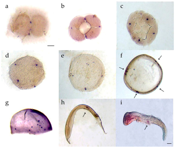
Figure 6
psdnd transcript localization during embryogenesis. Embryos were hybridized with antisense psdnd, stained by purple AP. (a) 2 cell stage: psdnd transcripts presented at first cleavage site; (b) 4 cell stage: psdnd positive signals revealed the spots where edges of second cleavage; (c) 32 cell stage; (d) morula stage: four dnd positive signals maintained until morula stage; (e) blastula stage: sister cells developed next to origin cells; (f) early gastrula stage; dnd transcripts increased and clustered at four spots of germ rings; (g) late gastrula stage; psdnd positive signals were move on to body axes; (h) early somite stage; (i) hatching larva: psdnd transcripts were aggregated and move on to future gonad location. Arrows indicate psdnd transcript expression. Scale bar: 100 μm.