IMAGE

FIGURE 3

ID
ZDB-IMAGE-210818-11
Source
Figures for Yang et al., 2021
Image
Figure Caption

FIGURE 3

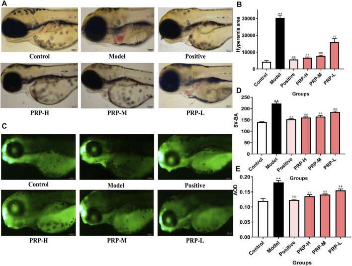
The effect of PRP on the heart development of zebrafish embryos following barium chloride treatment. (A) Morphological observations of the heart development of zebrafish embryos under a microscope, with the heart shown in the yellow box (X10). (B) The cardiac congestion area of zebrafish embryos was calculated. (C) The results of acridine orange staining. The apoptosis of zebrafish embryos cardiomyocytes observed under fluorescence microscopy is shown in the yellow box (X10). (D) The SV-BA distance was measured. (E) The average fluorescence density was calculated.All data are presented as means ± SD, n = 6. *p < 0.05, **p < 0.01, control versus model; #p < 0.05, ##p < 0.01, model versus positive, PRP-H, PRP-M, and PRP-L.

Acknowledgments
This image is the copyrighted work of the attributed author or publisher, and ZFIN has permission only to display this image to its users. Additional permissions should be obtained from the applicable author or publisher of the image. Full text @ Front Pharmacol