
Image
|
Figure Caption
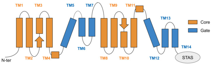
Figure 4 Schematic representation of the transmembrane domain of prestin, located between the N-terminal and the cytoplasmic, anti-sigma factor antagonist (STAS) domain. Helices are arranged according to a 7 transmembrane inverted repeat fold; they are conventionally indicated from 1 to 14, starting from the N-term, and are divided among the gate and core domains. The two arrows represent the short β-strands hosting the putative binding site of chloride ions.
Acknowledgments
This image is the copyrighted work of the attributed author or publisher, and
ZFIN has permission only to display this image to its users.
Additional permissions should be obtained from the applicable author or publisher of the image.
Full text @ Int. J. Mol. Sci.