
Figure 4
- ID
- ZDB-IMAGE-210801-99
- Publication
- Quadri et al., 2021 - Phosphorylation of H3-Thr3 by Haspin Is Required for Primary Cilia Regulation
- All Figures
- Figures for Quadri et al., 2021
|
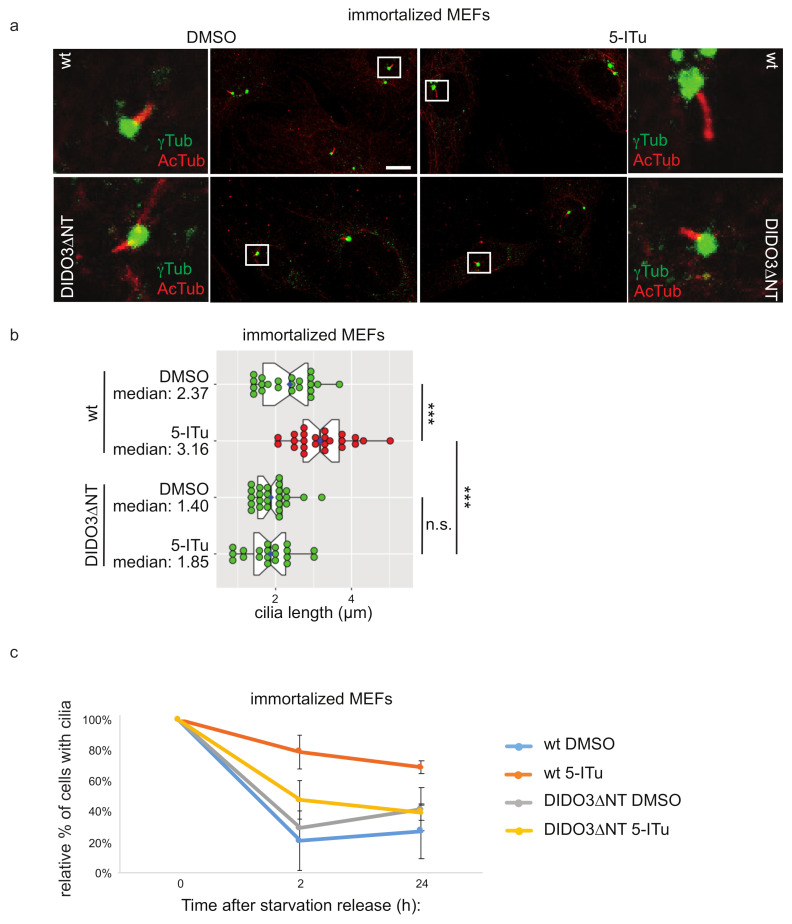
Figure 4 Displacement of Dido3 from the chromatin suppresses Haspin-dependent ciliary defects. Control or Dido3∆NT MEFs were driven in G0 by 48 h serum-starvation to induce ciliation and then treated overnight with DMSO or 10 nM 5-ITu ((a) and time 0 of (c)). At the end of the treatment, cells were incubated with a serum-containing medium to induce cilia resorption (in (c) for 2 and 24 h). Cilia length was measured by immunofluorescence analyzing acetylated tubulin and γ-tubulin (a). Representative images are shown in (a) (scale bar: 10 µm); cilia length is reported in graph (b); boxes include 50% of the data points, notch represent confidence interval (median ± 1.58 IQR/sqrt(n)). t-test was applied as a statistical measurement, n.s.; not significant, *** p < 0.005. Panel (c) shows cilia resorption kinetics represented as the normalized percentage of ciliated cells 2 h or 24 h after serum re-addition post-release. Error bars represent standard deviation.