IMAGE

Fig. 2

ID
ZDB-IMAGE-210801-34
Source
Figures for Wang et al., 2021
Image
Figure Caption

Fig. 2

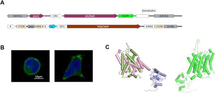
Frog prestin localized in the cell membrane. A Donor vector (upper one) designed for expression of ranaprestin-EGFP fusion protein and CRISPR/Cas9-mediated gene editing vector (lower one). B Membrane expression of rana prestin (rPres) in HEK293T cells was examined using confocal microscopy. The left one was a detached cell and the right one was an attached cell. C Predicted protein structure of rana prestin (left). The transmembrane domain holds two intertwined inverted repeats of seven TM segments. The N- and C-terminal halves of the TM domain are green and pink, respectively. The STAS domain is within the purple part. Predicted protein structure of gerbil prestin (right)

Acknowledgments
This image is the copyrighted work of the attributed author or publisher, and ZFIN has permission only to display this image to its users. Additional permissions should be obtained from the applicable author or publisher of the image. Full text @ BMC Genomics