
Fig. 2.
- ID
- ZDB-IMAGE-210801-17
- Publication
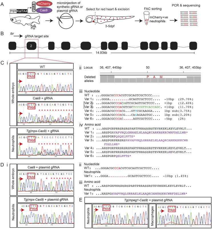
- Isiaku et al., 2021 - Transient, flexible gene editing in zebrafish neutrophils and macrophages for determination of cell-autonomous functions
- All Figures
- Figures for Isiaku et al., 2021
|
Fig. 2.