
|
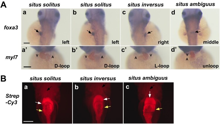
Figure 3. Situs solitus, situs inversus, and situs ambiguus in zebrafish visceral organ development (A) Whole mount double in situ hybridization reveals laterality of the developing liver (arrow) using a foxa3 RNA probe (purple) in the dorsal view of control (a) and experimental embryos (b–d) and the cardiac looping using the myocardial marker myl7 (brown) in the ventral view of control (a’) and experimental embryos (b’–d’). A, atrium; V, ventricle. Scale bars, 150 µm. (B) Ventral views of 6 dpf larvae stained with Streptavidin-Cy3 (Strep-Cy3) to visualize situs solitus (a), situs inversus (b), and situs ambiguus (c) of the heart (black arrow), liver (white arrow) and gut (yellow arrow). Scale bar, 150 µm.