
Fig. 3
- ID
- ZDB-IMAGE-210619-8
- Publication
- Zou et al., 2021 - In vivo imaging reveals mature Oligodendrocyte division in adult Zebrafish
- All Figures
- Figures for Zou et al., 2021
|
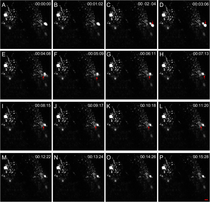
Fig. 3
Imaging proliferation of local oligodendrocytes in the transplanted optic nerve. Two olig2+ cells in the transplanted optic nerve project their processes to large areas, suggesting that they are complex oligodendrocytes. At 1 min 2 s, olig2+ cell division was not observed. At 2 min 4 s, a new olig2+ cell was divided from one olig2+ cell, and the whole progress was completed within 1 min. The newborn olig2+ cell migrated to the right at 3 min 6 s (descending arrow) and then migrated into the deeper space from 4 min 8 s to 11 min 20 s (ascending arrow). Finally, the cell migrated out of focus at 12 min 22 s. Each image has 25 optic slices, the interval is 1 μm, and the scale bar is 10 μm