IMAGE

Fig. 5

ID
ZDB-IMAGE-210619-10
Source
Figures for Zou et al., 2021
Image
Figure Caption

Fig. 5

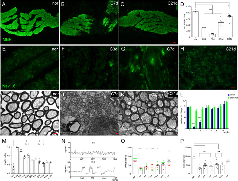
Visual function recovery after the completion of remyelination. a-c Qualitative results showing the restoration of MBP at different times after optic nerve injury. d Statistical results showing that the MBP intensity at the injury site was lowest at 7 dpi and gradually recovered in the following weeks. e-h Qualitative images showing Nav1.6 staining at different times after injury. At 7 dpi, Nav1.6 in the distal stump presented a linear pattern (g). i-k Qualitative images showing the myelin sheath at different times after optic nerve injury. Demyelination was completed at 7 dpi (j), but remyelination mostly occurred at 21 dpi (k). l Statistical results showing that the density of Ranvier nodes in the proximal region was not influenced by optic nerve injury, while it was greatly decreased in the distal region during the first two weeks and then returned to normal at 3 wpi. m Statistical results showing that the myelin index was gradually restored in the following weeks and completely finished at 9 wpi (p > 0.05). n Qualitative figures showing the nystagmus of normal and injured fish in the ocellanae OKR test. On the first day after optic nerve injury (down line), eye movement was voluntary (the large amplitude movements), and the nystagmus was much weaker in injuried fish than in normal fish (up line). o The preference of clockwise orientation in the injured eye was recovered at 3 wpi, and the OKR number was recovered at 4 wpi, indicated that the second-order visual function was restored. p The total amplitude of right eye nystagmus recovered, and the results indicated that fine visual function was restored at 6 wpi. The left side in A-C represents the proximal segment. Scale bar: 20 μm (a-c); 10 μm (e-h); 200 nm (i-k)

Acknowledgments
This image is the copyrighted work of the attributed author or publisher, and ZFIN has permission only to display this image to its users. Additional permissions should be obtained from the applicable author or publisher of the image. Full text @ Cell Regen (Lond)