
Fig. 2
- ID
- ZDB-IMAGE-210617-93
- Genes
- Antibodies
- Publication
- Peloggia et al., 2021 - Adaptive cell invasion maintains lateral line organ homeostasis in response to environmental changes
- All Figures
- Figures for Peloggia et al., 2021
|
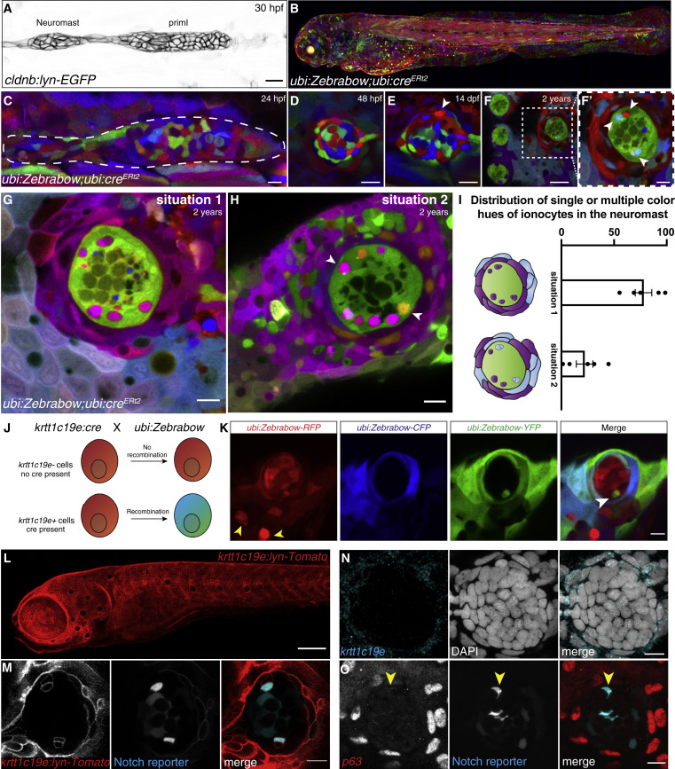
Fig. 2 (A) 30 hpf cldnb:lyn-GFP+ lateral line primordium and deposited neuromast. (B) Maximum intensity projection of a 3 dpf ubi:Zebrabow larva recombined during shield stage. (C–E) (C) 24 hpf primordium recombined for 15 min at 4 hpf. Neuromast in (C) is mosaic at 48 hpf (D) and 14 dpf (arrowhead indicates the new cell) (E). (F and F′) Neuromasts in 2-year-old fish recombined between 16–24 hpf have become clonal. Scale bar in (F), 30 μm. (G and H) (G) Z slices of 2-year-old recombined ubi:Zebrabow;ubi:creERt2 fish show Nm ionocyte pairs of the same color hue as skin cells surrounding the neuromast or (H) Nm ionocyte pairs of two different color hues (white arrows) also shared with surrounding skin cells. (I) Quantification of (G and H) (n = 5 fish, 194 neuromasts). Error bars indicate SEM. (J) Overview of lineage tracing experiments with ubi:Zebrabow and krtt1c19e:cre-MYC. (K) A 5 dpf ubi:Zebrabow;krtt1c19e:cre-MYC neuromast shows a pair of recombined cells (white arrowhead) in between red, unrecombined cells. Skin ionocytes differentiate before Cre expression and are not recombined (yellow arrowheads). (L) 5 dpf krtt1c19e:lyn-Tomato larva labels basal keratinocytes. Scale bar, 100 μm. (M) 5 dpf krtt1c19e:lyn-Tomato;tp1bglobin:EGFP neuromast. krtt1c19e channel was gamma adjusted. (N) krtt1c19e HCR and (O) TP63 immunohistochemistry does not label cells in the neuromast (Notch+ ionocytes indicated by red arrowhead). Scale bars, 10 μm, unless stated otherwise. See also Figure S2.
Reprinted from Developmental Cell, 56(9), Peloggia, J., Münch, D., Meneses-Giles, P., Romero-Carvajal, A., Lush, M.E., Lawson, N.D., McClain, M., Pan, Y.A., Piotrowski, T., Adaptive cell invasion maintains lateral line organ homeostasis in response to environmental changes, 1296-1312.e7, Copyright (2021) with permission from Elsevier. Full text @ Dev. Cell