IMAGE

Fig. 2

ID
ZDB-IMAGE-210617-8
Source
Figures for Hosseinzadeh et al., 2021
Image
Figure Caption

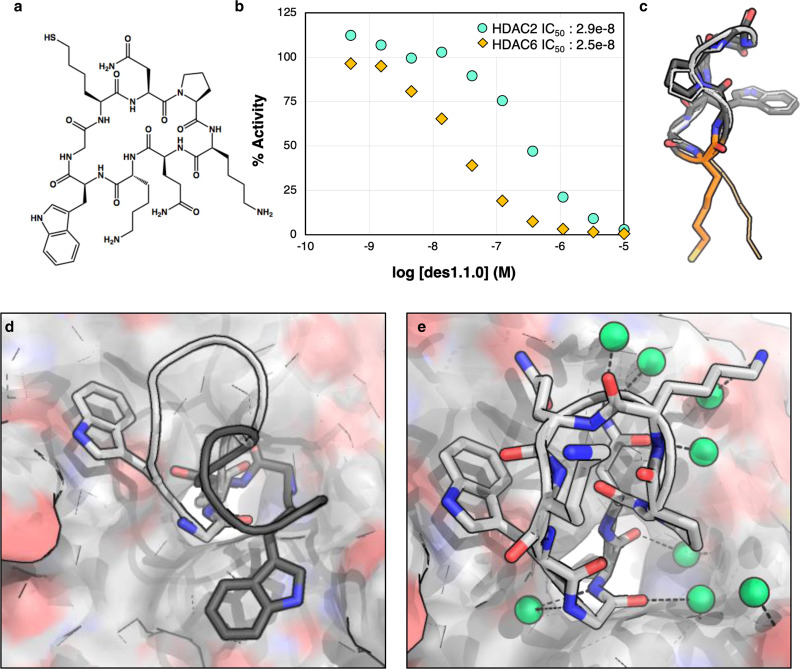
Fig. 2 Crystal structure of des1.1.0 has the designed monomer structure but adopts a different binding orientation.

a Chemical structure of des1.1.0. b des1.1.0 inhibits HDAC2 with an IC50 of 289 nM (Source Data are provided as a Source Data file). c Overlay of the designed peptide model des1.1.0 (dark gray) with the crystal structure (light gray). Some sidechains are removed for clarity. SHA anchor is shown in orange. d Overlay of des1.1.0 binding mode in original computational design (dark gray) and crystal structure (light gray) shows a clear rotation around SHA—Zn2+ axis in the binding pocket. e Crystal structure of des1.1.0 (PDB ID: 6WHO) shows several water-mediated interactions at the interface. Water molecules are shown as green spheres.

Acknowledgments
This image is the copyrighted work of the attributed author or publisher, and ZFIN has permission only to display this image to its users. Additional permissions should be obtained from the applicable author or publisher of the image. Full text @ Nat. Commun.