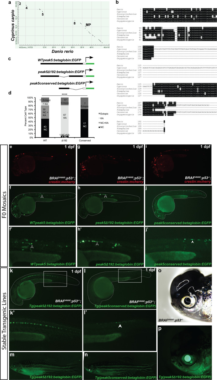
IMAGE

Fig. 4

ID
ZDB-IMAGE-210612-23
Source
Figures for Cunningham et al., 2021
Image
Figure Caption

Fig. 4 A conserved region of <italic>peak5</italic> is necessary for neural crest and melanoma activity.

a Dot-matrix view of the alignment of the carp (Cyprinus carpio) sox10 coding and surrounding noncoding genomic region of sox10 compared to the same genomic region in zebrafish (Danio rerio). Numbers indicate regions where peak sequences are conserved. MP minimal promoter. b Sequence alignment of the most conserved region of peak5 to members of the Cyprinidae family. c Schematic of plasmids used in the experiment. d Quantification of the percentage of categorizable EGFP + embryos exhibiting NC, KA, or both NC and KA labeling, or ectopic expression in screened WT peak5 (n = 91), peak5Δ192 (n = 89), and peak5_conserved (n = 24) injected embryos. NC neural crest. KA Kolmer–Agduhr neurons. ****p-value <0.0001, Chi-squared analysis. Injection of crestin:mCherry simply served as a positive injection control in this experiment. ecrestin:mch expression in an embryo injected with WT peak5 and crestin:mch. fWT peak5 is active in both neural crest and f′ KA neurons. gcrestin:mch expression in an embryo injected with peak5Δ192 and crestin:mch. hpeak5Δ192 is not active in NCCs, but h′ is active in KA neurons. icrestin:mch expression in an embryo injected with the conserved peak5 sequence and crestin:mch. j The conserved peak5 sequence is active in neural crest and j′ does not exhibit activity in KA neurons. Black arrowheads indicate KA neurons and white arrowheads indicate NCCs. kkTg(peak5Δ192:EGFP) embryos exhibit EGFP localization in KA neurons, but no overt localization in NCCs in the trunk is observed. ll′ Contrastingly, Tg(peak_conserved:EGFP) embryos do not have labeled KA neurons, but exhibit EGFP positive cells in the trunk of the embryo (white arrowhead). m Little EGFP localization is observed in Tg(peak5Δ192:EGFP) adult fish whereas n cells within the maxillary barbels (asterisk) of Tg(peak_conserved:EGFP) are EGFP positive. o, p The majority of tumors that develop in Tg(peak5Δ192:EGFP) are EGFP negative.

Acknowledgments
This image is the copyrighted work of the attributed author or publisher, and ZFIN has permission only to display this image to its users. Additional permissions should be obtained from the applicable author or publisher of the image. Full text @ Commun Biol