
|
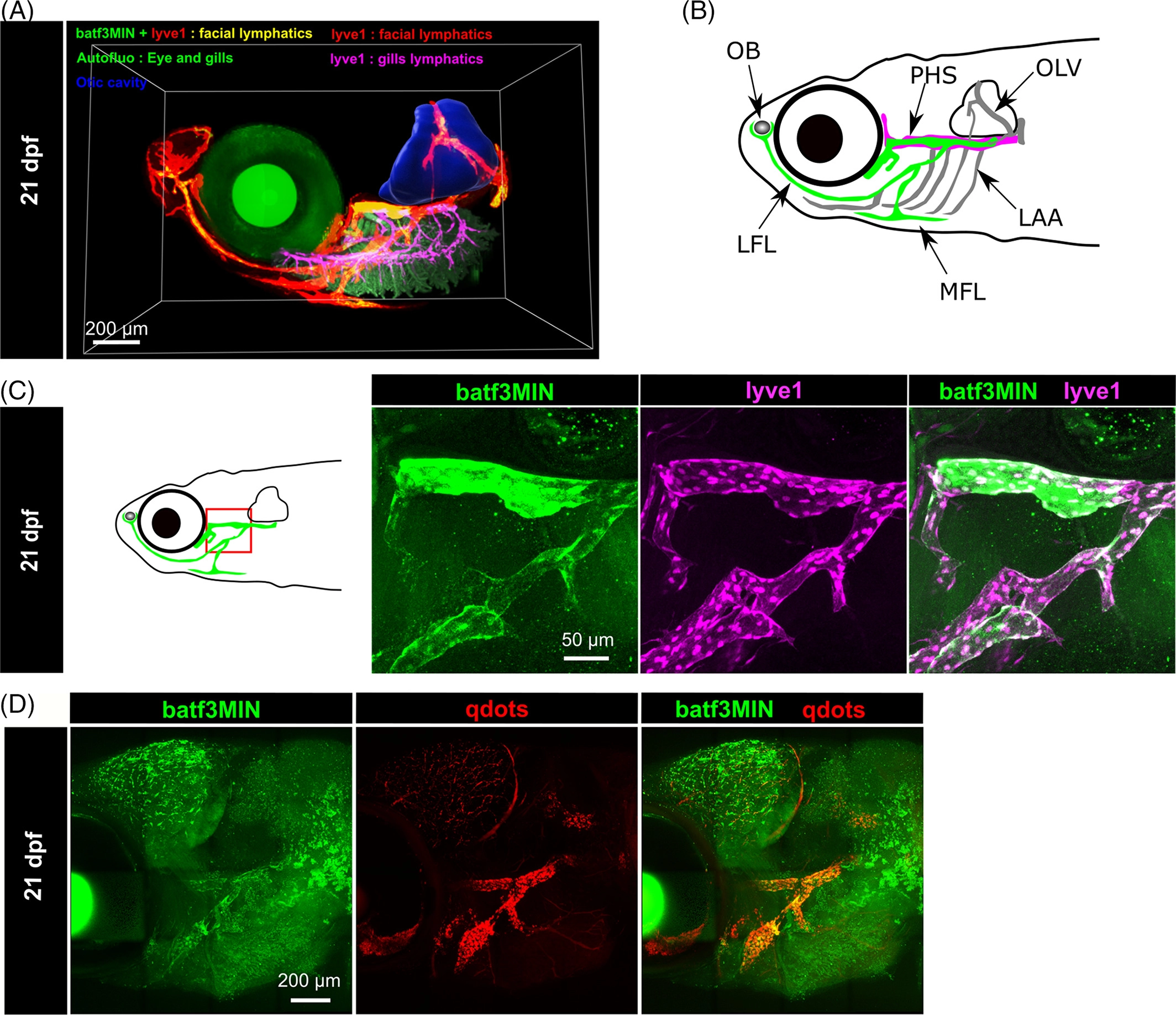
Fig. 3 In the Tg(batf3MIN:eGFP) reporter line, eGFP signal is restricted to a subset of facial lymphatic endothelial cell. A, Lateral view of segmented facial lymphatics in 21 dpf double Tg(batf3MIN:eGFP) x Tg(lyve1:dsRed2nz101) transgenic fish displayed in 3D. B, Schematic diagram of zebrafish facial lymphatics at 21 dpf. C, Schematic diagram of zebrafish head in lateral view. Red box indicated the region acquired. Maximum projection of a portion of facial lymphatics from double Tg(batf3MIN:eGFP) x Tg(lyve1:dsRed2nz101) transgenic fish at 21 dpf. D, Lymphangiogram of a 21dpf Tg(batf3MIN:eGFP) transgenic fish injected with qdots to visualize the facial lymphatics vessels. LFL, lateral facial lymphatics; MFL, medial facial lymphatics; LAA, lymphatic branchial arches; OLV, otolithic lymphatic vessel; PHS, primary head sinus; OB, olfactory bulbs