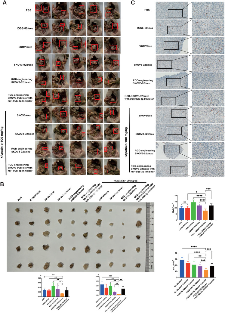
IMAGE

FIGURE 7

ID
ZDB-IMAGE-210601-20
Source
Figures for Wang et al., 2021
Image
Figure Caption

FIGURE 7

Tumor volume and angiogenesis were inhibited by engineered RGD‐SKOV3‐92b/exo alone or combined with Apatinib. (A) Gross anatomy images of nude mice in the peritoneal cavity. (B) Schematic representation of intraperitoneal xenografted tumors in different groups. Tumor size from each group was measured and weighed as represented. (C) Immunohistochemical staining of CD31 in intraperitoneal xenografted tumors (scale bar = 100 μm and 50 μm). Micro‐vessels density (MVD) was assessed by counting CD31‐positive cells (n = 5). Data are shown by at least three independent experiments. The Student's t‐test and nonparametric test were used to compare differences. *p < .05, **p < .01, ***p < .001, ****p < .0001

Acknowledgments
This image is the copyrighted work of the attributed author or publisher, and ZFIN has permission only to display this image to its users. Additional permissions should be obtained from the applicable author or publisher of the image. Full text @ Clin Transl Med