IMAGE

Figure 3

ID
ZDB-IMAGE-210528-7
Source
Figures for Wang et al., 2021
Image
Figure Caption

Figure 3

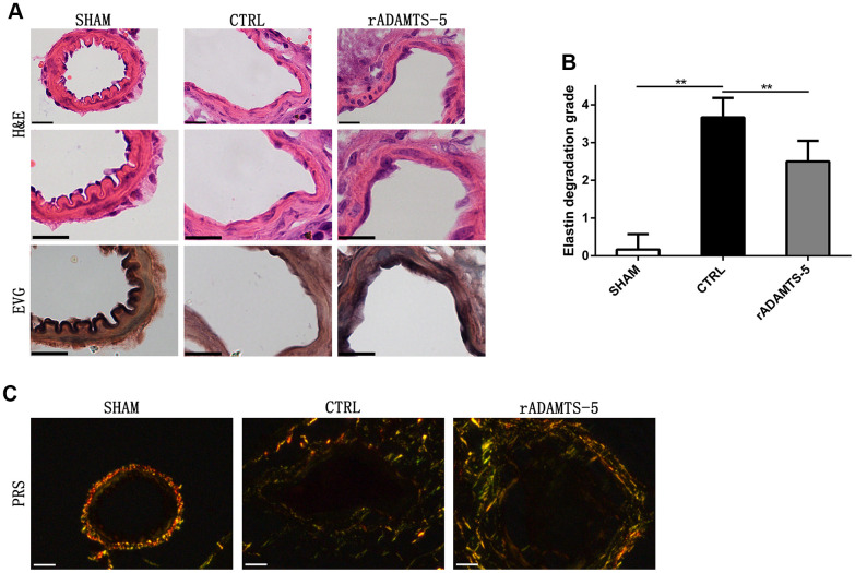
Recombinant protein ADAMTS-5 protected elastase-induced IA mice from vascular matrix degradation. (A) Representative hematoxylin and eosin (H&E) and elastin Verhoeff-Van Gieson (EVG)-stained section of cerebral arterial sections of sham mice, control (CTRL) IA mice, and recombinant protein ADAMTS-5-administered (rADAMTS-5) IA mice. Scale bar: 20 μm. (B) Quantification histogram showing elastin filament degradation. Arterial wall elastin filament degradation was evaluated based on the degree of degradation in elastin fibers (graded 0–4) as described in the Materials and Methods. **P<0.01. (C) Picrosirius red staining for collagen content within the arteries captured by polarization microscopy imaging from the sham mice, control IA mice, and recombinant protein ADAMTS-5-administered (rADAMTS-5) IA mice. Compared with the control group, the rADAMTS-5-administered group showed increased collagen birefringence under polarization. Scale bar: 20 μm.

Acknowledgments
This image is the copyrighted work of the attributed author or publisher, and ZFIN has permission only to display this image to its users. Additional permissions should be obtained from the applicable author or publisher of the image. Full text @ Aging (Albany NY)