
Figure 1
- ID
- ZDB-IMAGE-210519-74
- Genes
- Publication
- Knickmeyer et al., 2021 - BMP Signaling Interferes with Optic Chiasm Formation and Retinal Ganglion Cell Pathfinding in Zebrafish
- All Figures
- Figures for Knickmeyer et al., 2021
|
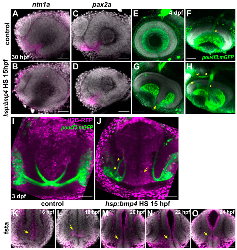
Figure 1 BMP signaling does not disrupt optic nerve head formation but prevents optic chiasm formation. (A–D) In situ hybridization of (A,B) ntn1a and (C,D) pax2a at 30 hpf in tg(hsp70l:bmp4) embryos and controls heat shocked at 15 hpf. DAPI counterstaining, sagittal view, nasal to the left. (E–H) Immunohistochemistry against GFP in tg(pou4f3:mGFP, hsp70l:bmp4) embryos and controls at 4 dpf. (E,F) Control eye, (F) tilted ventral projection. (G,H) Eye of an embryo with bmp4 induced at 15 hpf. RGC axons wrapping around the rim from the everted temporal retinal domain (arrow). (H) Tilted 3D projection of the eye depicted in (G). ONH is intact (asterisk), there is an aberrant nasal branch of the optic nerve (arrowheads). DAPI counterstaining, sagittal view, nasal to the left. (I,J) Live imaging of tg(pou4f3:mGFP, hsp70l:bmp4) embryos and controls at 3 dpf. (M) Control embryo. (N) Transgenic embryo with bmp4 induced at 15 hpf. RGCs project ipsilaterally. The right optic nerve possesses an additional nasal branch (arrowhead). There is no gap within the diencephalon (arrow). Transverse view. (K–O) In situ hybridization of fsta, midline expression (arrows) in the ventral prosencephalon of wild type embryos between 16–24 hpf. DAPI counterstaining, transverse view. All images are maximum intensity projections. Scale bars 50 µm.