
|
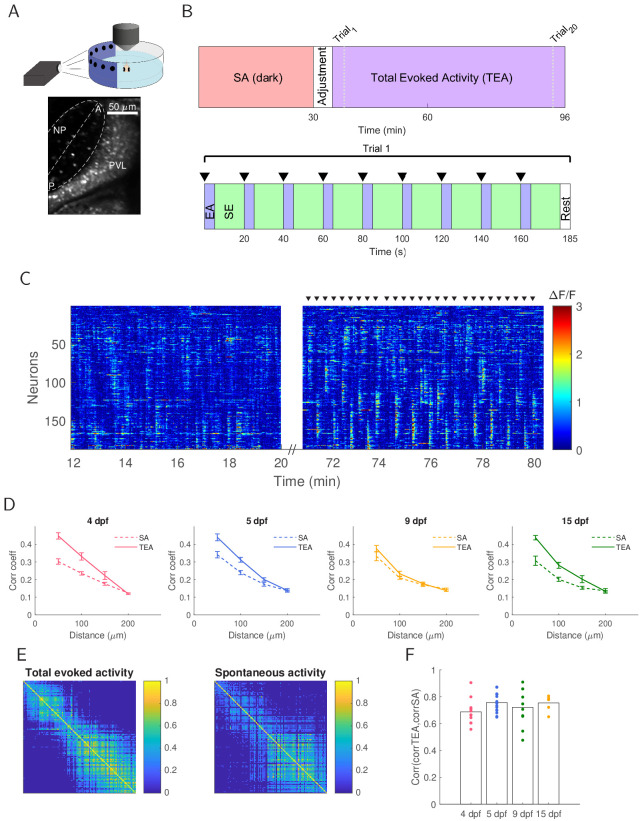
Figure 1 (A) Top: Larvae were embedded in agarose with one eye facing the projected image for two-photon calcium imaging. Bottom: The contralateral optic tectum (in this example 15 dpf) was imaged for 96.6 min. The neuropil (NP) contour of each fish was fitted with an ellipse (dashed line) with the major axis defining the tectal anterior-posterior axis (AP axis). Periventricular layer (PVL), NP, anterior (A) and posterior (P) ends of the tectum are indicated. (B) Experimental protocol. Tectal spontaneous activity (SA) in the dark was recorded after which fish were exposed to light and given 5 min to adjust. We then recorded evoked activity (TEA) in response to 20 trials of the stimulus set consisting of spots at positions 45°, 60°, 75°, 90°, 105°, 120°, 135°, 150°, 165° of the visual field (where 0° was defined as the body axis), presented in an order which maximised spatial separation within a trial. The inter-trial interval was 25 s. (C) Raster plot for an example 15 dpf fish showing concerted neural activity during 8 min of spontaneous activity in the dark and then during three cycles of stimulus presentation (stimulus onset is marked by black triangles). (D) Short-range pairwise correlation coefficients were higher for TEA compared to SA (4 dpf: p=10−4 for up to 50 µm, p=10−3 for 50–100 µm; 5 dpf: p=10−3 for up to 50 µm, p=10−3 for 50–100 µm; 15 dpf: p=10−3 for up to 50 µm, p=10−2 for 50–100 µm). (E) TEA and SA correlation matrices showed structural similarity (example shown is for a 15 dpf fish). Neurons were sorted by their position on the AP axis. (F) Correlation between TEA and SA correlation matrices does not change over development (one-way ANOVA, Bonferroni multiple comparison correction).