
|
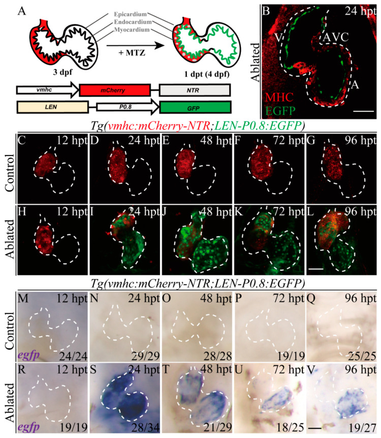
Figure 2 LEN is activated in the endocardium during ventricle regeneration. (A) Schematic diagrams of the transgenic constructs and ventricular ablation process of Tg(vmhc:mCherry-NTR; LEN-P0.8:EGFP) fish. (B) Confocal optical section image showed enhancer-directed fluorescence (green) in the endocardium of ablated Tg(vmhc:mCherry-NTR; LEN-P0.8:EGFP) hearts at 24 hpt. Red, MHC immunostaining by MF20. A, atrium; AVC, atrioventricular canal; MHC, myosin heavy chain; V, ventricle. (C–L) Confocal stack projections showed enhancer-directed fluorescence (green) dramatically increased in ablated hearts of Tg(vmhc:mCherry-NTR; LEN-P0.8:EGFP) fish compared to control hearts. (M–V) Whole-mount in situ hybridizations showed dynamic egfp expression in ablated hearts of Tg(vmhc:mCherry-NTR; LEN-P0.8:EGFP) compared to control hearts. Scale bars, (B–L) 50 μm, (M–V) 25 μm. Dashed lines outline the hearts. Numbers indicate the ratio of representative staining observed. dpf, days post fertilization; hpt, hours post treatment.