
IMAGE
Fig. 1
- ID
- ZDB-IMAGE-210426-16
- Publication
- Bieczynski et al., 2020 - Chemical effects on dye efflux activity in live zebrafish embryos and on zebrafish Abcb4 ATPase activity
- All Figures
- Figures for Bieczynski et al., 2020
Image
|
Figure Caption
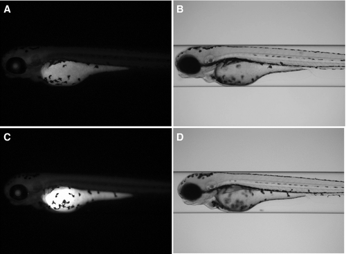
Fig. 1 Fluorescence (A, C) and bright‐field (B, D) images of live zebrafish embryos upon exposure to rhodamine B at 1 µm for 2 h. For imaging, the embryos were automatically positioned in lateral orientation in glass capillaries using the VAST system. (A, B) Solvent control (0.1% DMSO); (C, D) cyclosporin A (40 µm) treatment.
Acknowledgments
This image is the copyrighted work of the attributed author or publisher, and
ZFIN has permission only to display this image to its users.
Additional permissions should be obtained from the applicable author or publisher of the image.
Full text @ FEBS Lett.