
Image
|
Figure Caption
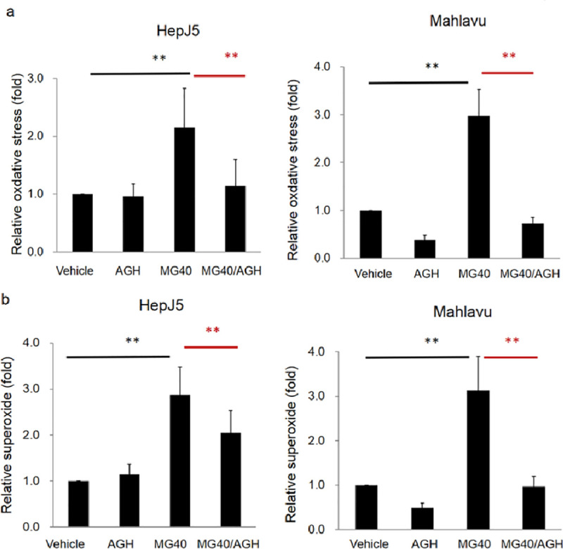
Fig 4 HepJ5 and Mahlavu cells were treated with AGH and then exposed to MG. Levels of ROS and superoxide were detected using specific fluorescence dyes. (a) Significant increase of the ROS level was found after MG treatment. The MG-induced ROS level was abolished in AGH pretreated HepJ5 and Mahlavu cells. (b) The superoxide level increased after exposure to MG and was abolished with AGH pretreatment. Data are presented as the mean±SD of three independent experiments in triplicate (** p<0.01).
Acknowledgments
This image is the copyrighted work of the attributed author or publisher, and
ZFIN has permission only to display this image to its users.
Additional permissions should be obtained from the applicable author or publisher of the image.
Full text @ PLoS One