
Image
|
Figure Caption
Fig 7
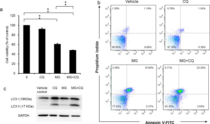
The cell viability in cells treated with MG for 24 h in the presence and absence of CQ for 16 h was determined using SRB assay (a). Apoptotic cells were quantified using Annexin V-FITC/PI staining and FACS analysis after 40 μg/ml MG treatment for 48h in the presence and absence of CQ for 16 h (b). The blockage of autophagy flux was confirmed by detecting the accumulation of LC3-II using western blot (c). Data are presented as the mean±SD of three independent experiments in triplicate, ** p<0.01).
Acknowledgments
This image is the copyrighted work of the attributed author or publisher, and
ZFIN has permission only to display this image to its users.
Additional permissions should be obtained from the applicable author or publisher of the image.
Full text @ PLoS One