
Image
|
Figure Caption
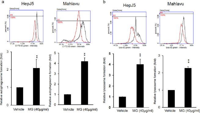
Fig 5 HepJ5 and Mahlavu cells were exposed to 40 μg/ml MG for 24 h. (a) Autophagosomes and (b) lysosomes were detected using specific dyes. The formation of autophagosomes and lysosomes increased after MG treatment compared to the vehicle. Data are presented as the mean±SD of three independent experiments in triplicate (** p<0.01).
Acknowledgments
This image is the copyrighted work of the attributed author or publisher, and
ZFIN has permission only to display this image to its users.
Additional permissions should be obtained from the applicable author or publisher of the image.
Full text @ PLoS One