
|
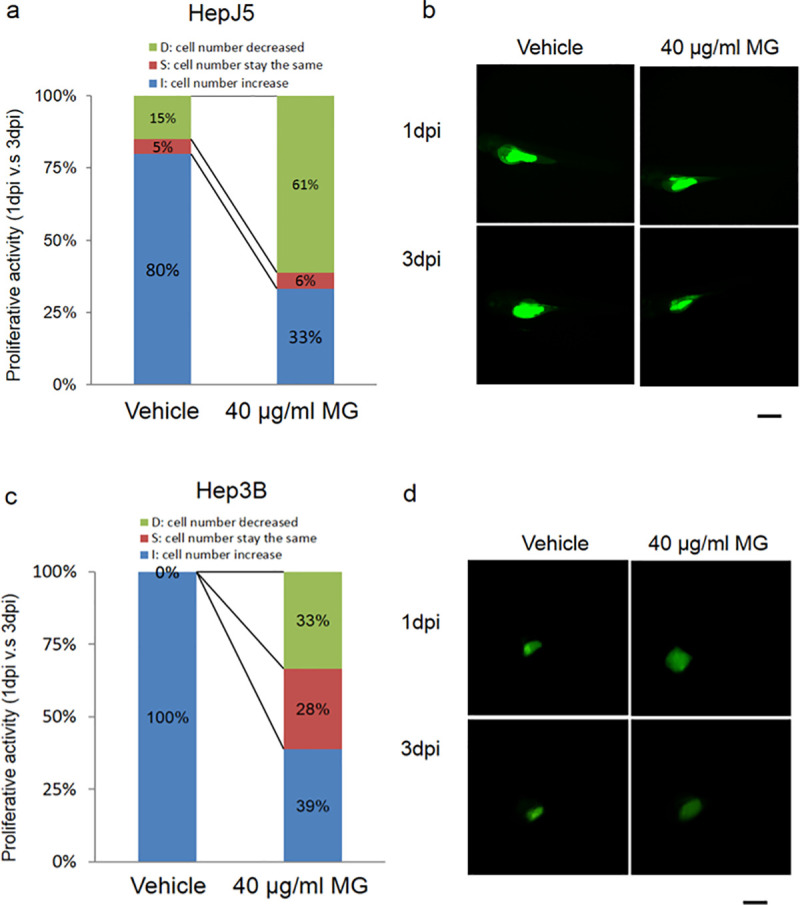
Fig 2 Zebrafish was used as the animal model for the xenotransplantation assay to determine the efficacy of MG treatment in hepatocellular carcinoma (HCC). Fluorescence labeled Hep3B and HepJ5 cells were implanted into an embryo yolk of the zebrafish, and then embryos were exposed to 40 μg/ml MG or dH2O as a vehicle control. Proliferative activities of the HCC cell lines in the embryos (n = 20 for each group) were compared by monitoring the fluorescence intensity on days 1 and 3 post-injection (1 and 3 dpi) of MG. (a and b) MG treatment reduced the increase in cell numbers in the embryo population (from 80% for the vehicle to 33% embryos respectively) in HepJ5 cells. A decrease in the fluorescence intensity was shown after 3 days in Hep3B cells with 40 μg/ml MG treatment (c and d). In the Hep3B cell line, the increase in cell numbers in the embryo population decreased from 100% (vehicle) to 39% (40 μg/ml) within 20 embryos. Treatment with 40 μg/ml MG dramatically decreased the fluorescence intensity in HCC cells compared to the vehicle. Scale bare was 1 mm.