IMAGE

Fig. 1

ID
ZDB-IMAGE-210325-39
Source
Figures for Fan et al., 2021
Image
Figure Caption

Fig. 1

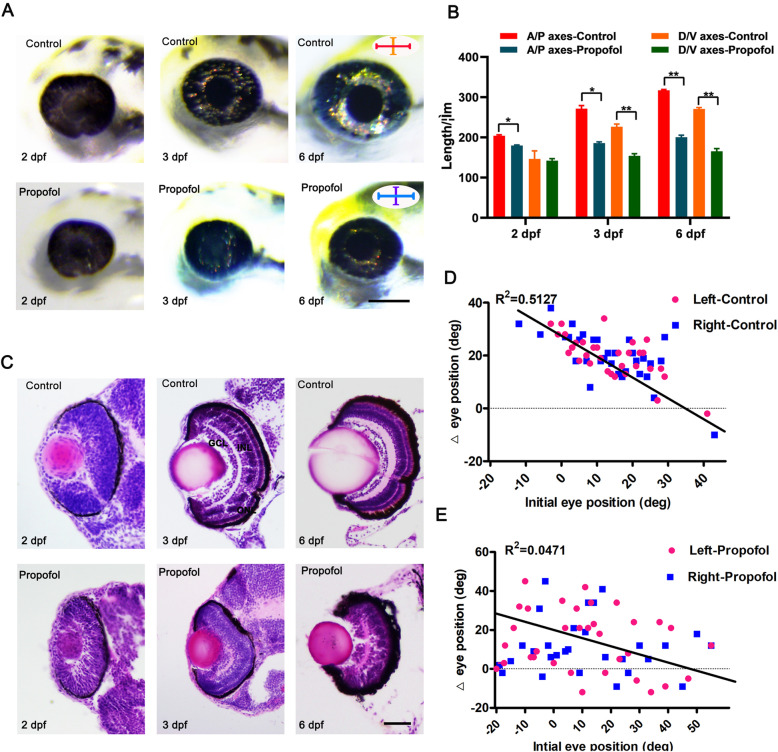
Propofol treatment results in microphthalmia and defects in retinal lamination and function. a Compared with control embryos, propofol-treated embryos (5 μg/ml propofol) at 2, 3, and 6 days postfertilization (dpf) display a reduction in eye size. b Measurements of absolute eye size along the anteroposterior (A/P) and dorsoventral (D/V) axes at 2, 3, and 6 dpf. n = 12. Error bars indicate the standard error of the mean. c Transverse histological sections of retinas of control and propofol-treated embryos (5 μg/ml propofol) at 2, 3, and 6 dpf. Eyes of propofol-treated embryos (5 μg/ml propofol) are smaller than control eyes, and the ganglion cell layer, inner nuclear layer, and outer nuclear layer are not morphologically identifiable in the retinas of propofol-treated embryos at 3 dpf. The dorsal side is up in all images. n = 7. d, e In response to prey, the extent of eye rotation is linearly associated with the initial position of the eye in control larvae at 6 dpf; in propofol-treated larvae (5 μg/ml propofol), there is no convergent eye movement. n = 35. a Scale bar, 100 μm. c Scale bar, 35 μm

Acknowledgments
This image is the copyrighted work of the attributed author or publisher, and ZFIN has permission only to display this image to its users. Additional permissions should be obtained from the applicable author or publisher of the image. Full text @ Stem Cell Res. Ther.