
Fig. 1
- ID
- ZDB-IMAGE-210316-7
- Publication
- Bertozzi et al., 2020 - Is zebrafish heart regeneration "complete"? Lineage-restricted cardiomyocytes proliferate to pre-injury numbers but some fail to differentiate in fibrotic hearts
- All Figures
- Figures for Bertozzi et al., 2020
|
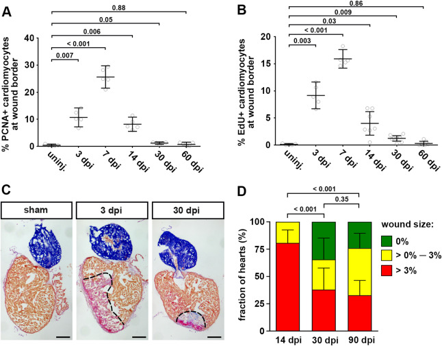
Fig. 1 Fig. 1. Cardiomyocyte cell cycle activity drops to low levels prior to complete morphological regeneration. (A) The fraction of PCNA+ EGFP+ cardiomyocytes in myl7:EGFPtwu34 fish within 150 μm of the wound border peaks at 7 dpi and drops to levels barely above baseline by 30 dpi. uninj, uninjured. Error bars represent CI 95%. n = 5 hearts for each group. One-way ANOVA + Dunnett’s multiple comparisons test. Numbers indicate p-value. (B) Fraction of EdU+ EGFP+ cardiomyocytes in myl7:EGFPtwu34 fish. For each time point, a single intraperitoneal injection of EdU was performed 24 h prior to heart extraction. n (hearts) = 5 (uninjured), 5 (3 dpi), 5 (7 dpi), 7 (14 dpi), 8 (30 dpi), 7 (60 dpi). One-way ANOVA + Dunnett’s multiple comparisons test. (C) Many hearts contain wound tissue detected by Acid Fuchsin Orange G (AFOG) staining at 30 dpi. Red: fibrin, blue: collagen, brown: myocardium. Scale bars, 100 μm. (D) Percentage of hearts without residual wounds (= 0% of ventricle area), small wounds (between 0% and 3%) or bigger wounds (>3%) at 14, 30 and 90 dpi. n = 31 (14 dpi), 29 (30 dpi), 58 (90 dpi). Chi-square test.
Reprinted from Developmental Biology, 471, Bertozzi, A., Wu, C.C., Nguyen, P.D., Vasudevarao, M.D., Mulaw, M.A., Koopman, C.D., de Boer, T.P., Bakkers, J., Weidinger, G., Is zebrafish heart regeneration "complete"? Lineage-restricted cardiomyocytes proliferate to pre-injury numbers but some fail to differentiate in fibrotic hearts, 106-118, Copyright (2020) with permission from Elsevier. Full text @ Dev. Biol.