IMAGE

Figure 7

ID
ZDB-IMAGE-210310-10
Source
Figures for Iscan et al., 2021
Image
Figure Caption

Figure 7

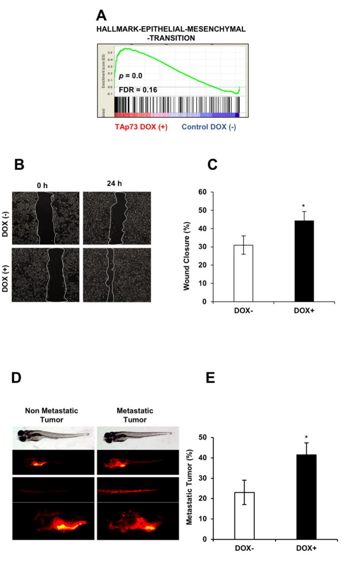
TAp73β upregulates epithelial-mesenchymal transition hallmark genes and stimulates metastatic abilities of hepatocellular carcinoma cells. (A) Genes involved in epithelial-mesenchymal transition in TAp73β induced cells. (B) Wound-healing assay was performed to examine the migration rate of Hep3B cells under TAp73β induction. Images were captured at 0 and 24 h following the initial scratch. (C) Migration rates were quantified by measuring three different wound areas using ImageJ software, columns and error bars indicate mean ± SD from triplicate experiments. The graph is representative of three independent experiments. (D) This figure illustrates metastatic (right panel) and non-metastatic (left panel) tumors after 4 days grown in zebrafish larvae. Bright field picture of larvae was shown in the top level the bottom level pictures are fluorescent images of tumors in same animals. Fluorescent pictures in the middle shows metastatic tumor cells disseminated into tail region (right) as opposed to non-metastatic tumor cells (left). Hep3BTAp73β cells were treated with doxycycline 1 µg/mL for 72 h, stained with Dil and inoculated into the 2 dpf zebrafish embryos. Bright-field and fluorescent images of zebrafish were captured for non-metastatic and metastatic tumors. The number of embryos with metastasis was counted at day 4 dpi (days post-injection) (E). Three separate experiments were performed. Error bars, ± SD (number of zebrafish embryo, n = 30). * p < 0.05.

Figure Data
Acknowledgments
This image is the copyrighted work of the attributed author or publisher, and ZFIN has permission only to display this image to its users. Additional permissions should be obtained from the applicable author or publisher of the image. Full text @ Cancers