IMAGE

Figure 4

ID
ZDB-IMAGE-210305-6
Source
Figures for Lämmle et al., 2020
Image
Figure Caption

Figure 4

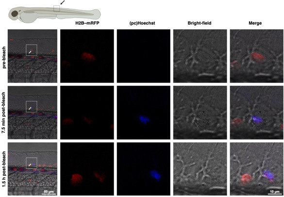
In vivo uncaging of pcHoechst with optimized light intensity at single‐cell resolution. H2B‐mRFP (red) injected zebrafish embryos were incubated with 100 μM pcHoechst in the dark overnight and activated by using a 405 nm UV laser at 0.07 mW and at 74 hpf. The arrow indicates a nucleus targeted with two bleach points. A pre‐bleach image was recorded prior to illumination, post bleach images 7.5 min and 1.5 h after bleaching. The white rectangles depict magnified areas. Uncaged pcHoechst (blue) can be observed 7.5 min and 1.5 h after UV illumination and colocalizes with H2B‐mRFP (red) in a single nucleus. Representative images of two experiments with the same intensity, and ten experiments in total.

Acknowledgments
This image is the copyrighted work of the attributed author or publisher, and ZFIN has permission only to display this image to its users. Additional permissions should be obtained from the applicable author or publisher of the image. Full text @ Chembiochem