IMAGE

Fig 1

ID
ZDB-IMAGE-210304-1
Source
Figures for Lu et al., 2021
Image
Figure Caption

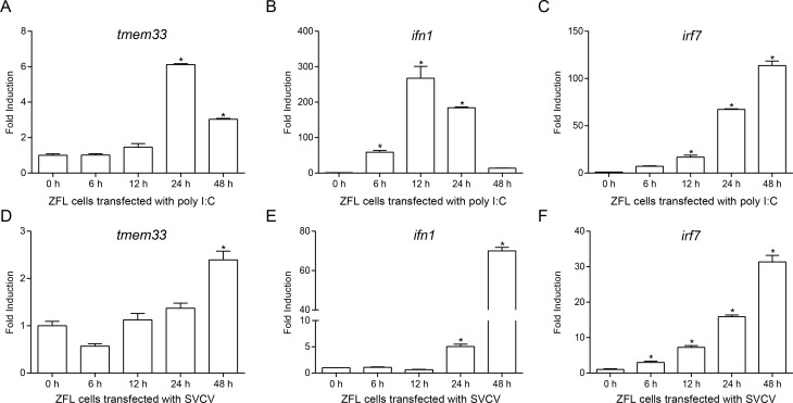
Fig 1 TMEM33 is stimulated by virus infection.

(A-F) qPCR detection of the transcriptional levels of tmem33, ifn1, and irf7 on stimulation. ZFL cells seeded on 6-well plates overnight and transfected with poly I:C (1 μg/ml) (A-C) or stimulated with SVCV (MOI = 1) (D-F). At the time points 6, 12, 24, and 48 h, total RNAs were extracted for further qPCR assays. β-actin was used as an internal control for normalization and the relative expression is represented as fold induction relative to the expression level in control cells (set to 1). Data were expressed as mean ± SEM, n = 3. Asterisks indicate significant differences from control (*, p < 0.05).

Acknowledgments
This image is the copyrighted work of the attributed author or publisher, and ZFIN has permission only to display this image to its users. Additional permissions should be obtained from the applicable author or publisher of the image. Full text @ PLoS Pathog.