
|
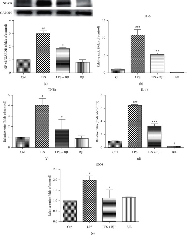
Figure 3 BJL partially inhibited LPS-induced upregulations of NF-κB, iNOS, and proinflammatory cytokines in RAW 264.7 cells. RAW 264.7 cells were treated LPS (0.3 μg/ml) with or without BJL (30 μg/ml) for 24 h. (a) The intracellular protein level of NF-κB was tested by western blotting using the specific primary antibody. (b–e) The mRNA expression levels of TNF-α, IL-6, IL-1β, and iNOS were detected by real-time PCR. Data were presented as the folds of the control group. Results were means ± S.E.M. of more than 3 independent experiments. p<0.055, p<0.01, and p<0.001 versus the control group. *p<0.05, **p<0.01, and ***p<0.001 versus the LPS-treated group.