
Fig. 2
- ID
- ZDB-IMAGE-210217-41
- Publication
- Okada et al., 2020 - The second pharyngeal pouch is generated by dynamic remodeling of endodermal epithelium in zebrafish
- All Figures
- Figures for Okada et al., 2020
|
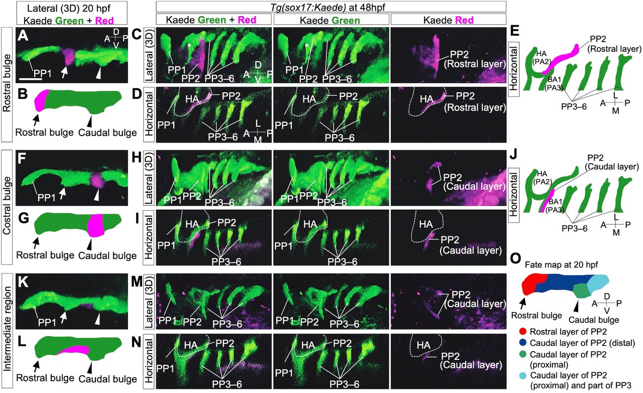
Fig. 2 Lineage tracing of endodermal cells in Tg(sox17:Kaede) zebrafish embryos by photoconversion. (A-E) Cells of the rostral bulge (arrows) were marked at 20 hpf (A,B). At 48 hpf, cells of the rostral bulge contributed to the large area of the rostral portion of PP2 (C-E). (F-J) The cells of the caudal bulge (arrowheads) were marked at 20 hpf (F,G). At 48 hpf, the descendant cells contributed to the caudal region of PP2 rather proximally (H-J). (K-N) The cells of the intermediate domain of a putative PP2 (between the rostral and caudal bulges) were marked (K,L). At 48 hpf, these descendants composed the dorsally and ventrally distant area in the caudal part of PP2 (M,N). (O) Overview of the cell fate of future PP2 endoderm at 20 hpf. Cell fates were examined in various regions of the presumptive PP2 endoderm by photoconversion (n=29, A-N and Fig. S1); these are summarized, showing the dynamic reorganization of the endoderm forming PP2. A, anterior; BA, branchial arch; D, dorsal; HA, hyoid arch; L, lateral; M, medial; P, posterior; PP1-6, the first to sixth pharyngeal pouches; V, ventral. The asterisk indicates a blood vessel. The positions of the HA (dashed lines) were identified by the surrounding sox17-positive endoderm. Scale bar: 50 μm.