
|
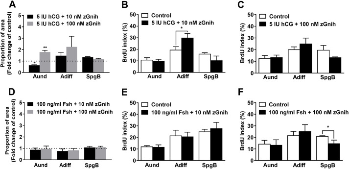
Fig. 7 Fig. 7. (A and D) Proportion of type A undifferentiated spermatogonia (Aund), type A differentiated (Adiff) and type B spermatogonia (SpgB) were determined in the section surface area within the spermatogenic cysts in the absence (dotted line) or following various treatments as shown. Bars show the mean ± SEM (t-test; n = 5) and asterisks indicate significant difference with respect to basal control (dotted line), using Student paired t-test (n = 5). B, C, E and F show BrdU-incorporation during the last 6 h of 7-days testis culture. BrdU positive germ cells: Aund, Adiff and SpgB were counted in the testes. BrdU positive germ cells: Aund, Adiff and SpgB were counted in the testes following various treatments shown. Bars shows mean ± SEM (n = 5). Each treatment was compared with its own (individual) control (contralateral testis). Asterisks over connected line denote statistical significance between the control and treatment group, using Student's T-test.
Reprinted from Molecular and Cellular Endocrinology, 520, Fallah, H.P., Rodrigues, M.S., Zanardini, M., Nóbrega, R.H., Habibi, H.R., Effects of gonadotropin-inhibitory hormone on early and late stages of spermatogenesis in ex-vivo culture of zebrafish testis, 111087, Copyright (2020) with permission from Elsevier. Full text @ Mol. Cell. Endocrinol.