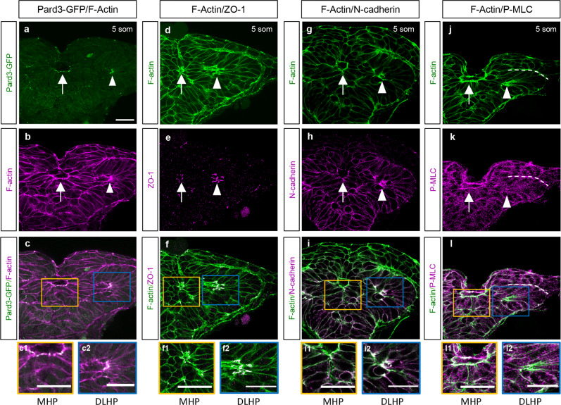
a–l Transverse sections through the ANP at the 5 som stage. Embryos double-labeled with Pard3-GFP (green) and phalloidin (F-actin, magenta) (a–c2); ZO-1 (magenta) and F-actin (green) (d–f2); N-cadherin (magenta) and F-actin (green) (g–i2); and with anti-P-MLC (magenta) and F-actin (green) (j–l2). c, f, i, l Magenta and green channel overlay. Insets show higher magnification images of the MHP (yellow boxes, c1, f1, i1, l1) and DLHP (blue box, c2, f2, i2, l2). Annotations: arrow = MHP; arrowhead = DLHP; dotted line in j–l = interface between the neuroectoderm and non-neural ectodermlayers of the neural folds; MHP medial hingepoint, DLHP dorsolateral hingepoint. Scale bars: 25 μm.
Acknowledgments
This image is the copyrighted work of the attributed author or publisher, and
ZFIN has permission only to display this image to its users.
Additional permissions should be obtained from the applicable author or publisher of the image.
Full text @ Commun Biol
Your Input Welcome
Thank you for submitting comments. Your input has been emailed to ZFIN curators who may contact you if
additional information is required.
Oops. Something went wrong. Please try again later.