IMAGE

Figure 6—figure supplement 4.

ID
ZDB-IMAGE-210204-97
Publication
Parab et al., 2021 - Endothelial cell type-specific molecular requirements for angiogenesis drive fenestrated vessel development in the brain
All Figures
Figures for Parab et al., 2021
Image
Figure Caption

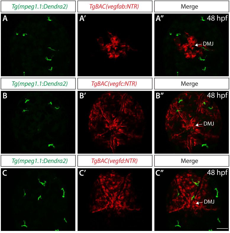
Figure 6—figure supplement 4. Co-localization analyses in <italic>TgBAC(vegfab:NTR)</italic>, <italic>TgBAC(vegfc:NTR)</italic>, and <italic>TgBAC(vegfd:NTR)</italic> embryos carrying the <italic xmlns:xlink='http://www.w3.org/1999/xlink'>Tg(mpeg1.1:Dendra2)</italic> transgene.

(A–A") Confocal maximum projection images of the dorsal views of a 48 hpf Tg(mpeg1.1:Dendra2);TgBAC(vegfab:NTR) embryo, showing non-overlapping distribution of EGFP+ and NTR-mCherry+ meningeal cells (n = 16 embryos; 89 Tg(mpeg1.1:Dendra2)+ cells examined; 0 cells co-localized with TgBAC(vegfab:NTR)+ cells). (B–B") Confocal maximum projection images of the dorsal views of a 48 hpf Tg(mpeg1.1:Dendra2);TgBAC(vegfc:NTR) embryo, showing non-overlapping distribution of EGFP+ and NTR-mCherry+ meningeal cells (n = 17 embryos; 95 Tg(mpeg1.1:Dendra2)+ cells examined; 0 cells co-localized with TgBAC(vegfc:NTR)+ cells). (C–C") Confocal maximum projection images of the dorsal views of a 48 hpf Tg(mpeg1.1:Dendra2);TgBAC(vegfd:NTR) embryo, showing non-overlapping distribution of EGFP+ and NTR-mCherry+ meningeal cells (n = 8 embryos; 39 Tg(mpeg1.1:Dendra2)+ cells examined; 0 cells co-localized with TgBAC(vegfd:NTR)+ cells). Scale bar: 50 μm.

Acknowledgments
This image is the copyrighted work of the attributed author or publisher, and ZFIN has permission only to display this image to its users. Additional permissions should be obtained from the applicable author or publisher of the image. Full text @ Elife