IMAGE

Figure 5

ID
ZDB-IMAGE-210201-11
Source
Figures for Babi? et al., 2021
Image
Figure Caption

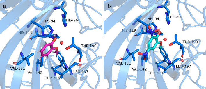
Figure 5

Representation of the interactions between (a) GUA and (b) 4,6DNG with the active site D. rerio CA II. Structure of D. rerio CA II (UniProt entry: Q92051) is modeled by I-TASSER, coordinates for Zn cation, and active site water molecule (oxygen atom in red) were taken from the structure of human CA deposited under PDB ID 1fql, whereas docking pose for GUA was obtained by SwissDock server. The same pose was obtained by AutoDock vina. Representation was made in PyMOL software (https://pymol.org/2/).

Acknowledgments
This image is the copyrighted work of the attributed author or publisher, and ZFIN has permission only to display this image to its users. Additional permissions should be obtained from the applicable author or publisher of the image. Full text @ Sci. Rep.