
IMAGE
Figure 2
- ID
- ZDB-IMAGE-210128-10
- Publication
- Warchol et al., 2021 - Macrophages Respond Rapidly to Ototoxic Injury of Lateral Line Hair Cells but Are Not Required for Hair Cell Regeneration
- All Figures
- Figures for Warchol et al., 2021
Image
|
Figure Caption
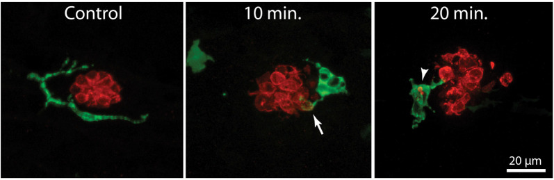
Figure 2
Macrophage response to neomycin ototoxicity. Images are maximum-intensity projections of confocal
Acknowledgments
This image is the copyrighted work of the attributed author or publisher, and
ZFIN has permission only to display this image to its users.
Additional permissions should be obtained from the applicable author or publisher of the image.
Full text @ Front. Cell. Neurosci.