
Figure 1
- ID
- ZDB-IMAGE-210121-18
- Publication
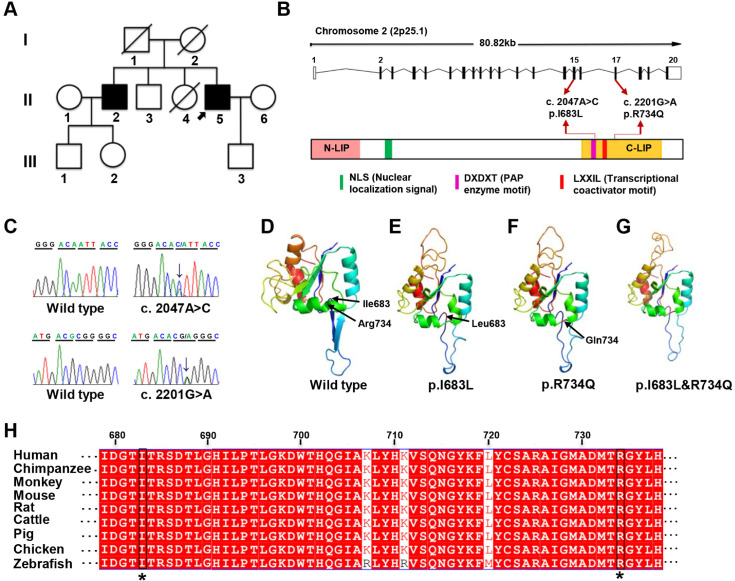
- Lu et al., 2021 - Lipin 1 deficiency causes adult-onset myasthenia with motor neuron dysfunction in humans and neuromuscular junction defects in zebrafish
- All Figures
- Figures for Lu et al., 2021
|
Figure 1