
Figure 5
- ID
- ZDB-IMAGE-210117-23
- Publication
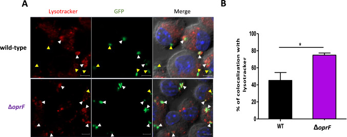
- Moussouni et al., 2021 - Pseudomonas aeruginosa OprF plays a role in resistance to macrophage clearance during acute infection
- All Figures
- Figures for Moussouni et al., 2021
|
Figure 5
Colocalization of