IMAGE

Figure 3

ID
ZDB-IMAGE-210108-23
Genes
Source
Figures for Hahn et al., 2020
Image
Figure Caption

Figure 3 Early expression of p75 NTR (<italic>ngfrb</italic>) is seen in two sets of cranial ganglia using whole mount in situ hybridization at 24 hpf.

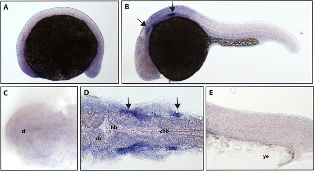
(A and C) Expression of ngfrb was not detected at 16.5 hpf and the staining that can be seen was non-specific (Fig. S2). (B, D and E) At 24 hpf, expression of ngfrb was seen in two sets of cranial ganglia (arrows). (A, B and E) Lateral view. (C and D) Dorsal view. All embryos are oriented with the head to the left. d, marks the diencephalon, cb, marks the cerebellum, hb, marks the hindbrain, chb, marks the caudal hindbrain and ye, marks the yolk extension. Data are representative of the results from two rounds of in situ hybridization with at least 25 embryos per round.

Figure Data
Acknowledgments
This image is the copyrighted work of the attributed author or publisher, and ZFIN has permission only to display this image to its users. Additional permissions should be obtained from the applicable author or publisher of the image. Full text @ Peer J.