IMAGE

Figure 7

ID
ZDB-IMAGE-201229-20
Source
Figures for Tonon et al., 2020
Image
Figure Caption

Figure 7

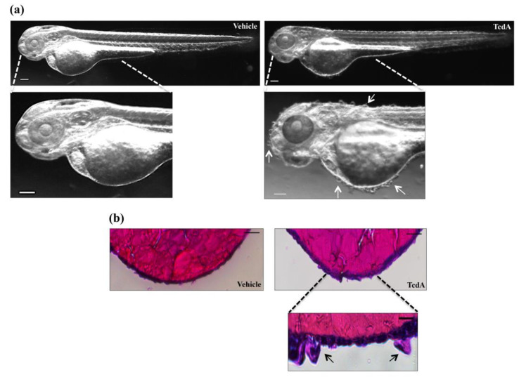
TcdA administration induced skin alteration in zebrafish embryos. (a) Representative images of 2.5 µg/mL TcdA- or vehicle-treated embryos. Images were acquired with a Leica S8AP0 stereo microscope. Scale bar = 100 µm. (b) Hematoxylin and eosin staining is performed on 2.5 µg/mL TcdA- or vehicle-treated embryos to evaluate skin structure. Scale bar = 100 µm (upper panel). A magnification of the hypertrophic cell of the epidemical layer is taken from TcdA-treated embryo (lower panel; scale bar = 10 µm). Images were acquired with a Leica S8AP0 stereo microscope.

Acknowledgments
This image is the copyrighted work of the attributed author or publisher, and ZFIN has permission only to display this image to its users. Additional permissions should be obtained from the applicable author or publisher of the image. Full text @ Cells