Image
Figure Caption

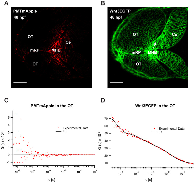
Figure 3—figure supplement 1. Fluorescence correlation spectroscopy (FCS) measurements of Wnt3EGFP and PMTmApple in the Wnt3 target regions at 48 hpf.

Expression of (A) PMTmApple and (B) Wnt3EGFP in Tg(−4.0wnt3:Wnt3EGFP) × Tg(−4.0wnt3:PMTmApple) double transgenics at 48 hpf. Representative autocorrelation function (ACF) for (C) PMTmApple and (D) Wnt3EGFP in the OT at 48 hpf. No ACFs were obtained for PMTmAPple in the target regions indicating an absence of mApple signal in these regions. Ce, cerebellum; MHB, midbrain–hindbrain boundary; OT, optic tectum. Images orientation: anterior to the left. Scale bar 30 μm.

Acknowledgments
This image is the copyrighted work of the attributed author or publisher, and ZFIN has permission only to display this image to its users. Additional permissions should be obtained from the applicable author or publisher of the image. Full text @ Elife