IMAGE

Fig. 4

ID
ZDB-IMAGE-201121-34
Source
Figures for Kondrychyn et al., 2020
Image
Figure Caption

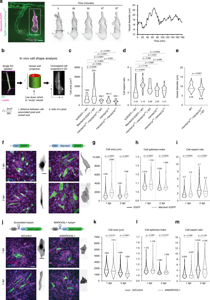
Fig. 4 Marcksl1 expression level controls endothelial cell size and shape.

a Live imaging of an ISV composed of EC with ectopic expression of Marcksl1b from 48 hpf reveals changes in cell shape and fluctuation in vessel diameter. (n = 5 embryos, three independent experiments). Dashed line, region of vessel measured. bd Overexpression of Marcksl1 increases EC size while depletion of marcksl1b or marcksl1a and marcksl1b decreases EC size in vivo. Methodology to perform in vivo single-cell shape analysis (b). Quantification of cell area (c) and cell aspect ratio (d) of lynEGFP- or Marcksl1b-EGFP-expressing ECs surrounded by wildtype (WT) cells, marcksl1rk23;marcksl1brk24 ECs transplanted into WT embryo and ECs of marcksl1brk24 or marcksl1rk23;marcksl1brk24 embryos at 2 dpf. e Diameter of ISVs composed of single marcksl1rk23;marcksl1brk24 EC transplanted into WT embryo at 2 dpf (n = 10 vessels, n = 5 independent transplantations). fm Overexpression and knockdown of Marcksl1 increases and decreases, respectively, HUVEC size (n = 3 independent transfections). Maximum intensity projection of confocal z-stacks of control (EGFP) and Marcksl1-overexpressing (Marcksl1-EGFP) HUVECs 1 and 2 days post transfection (dpt, f). Quantification of cell area (g), cell spikiness index (h) and cell aspect ratio (i, EGFP: n = 423/363 cells at 1/2 dpt; Marcksl1-EGFP: n = 358/298 cells at 1/2 dpt). Maximum intensity projection of confocal z-stacks of control and MARCKSL1 knockdown HUVECs stained for DAPI and phalloidin at 1 and 2 dpt (j). Plasmid expressing U6 promoter-driven MARCKSL1 shRNA was used with CMV promoter-driven EGFP-CAAX expression as an internal marker. Plasmid expressing scrambled shRNA was used as a control. Quantification of cell area (k), cell spikiness index (l) and cell aspect ratio (m) after MARCKLS1 knockdown (shControl, n = 223/278 at 1/2 dpt; shMARCKSL1, n = 474/342 at 1/2 dpt). Statistical significance was assessed by ordinary one-way ANOVA with Sidak’s multiple comparisons test (c, d) and two-tailed unpaired t-test (e, gi, km). Violin plots in (ce, gi and km) represent the entire range of values, dotted lines indicate first and third quartiles, center lines are median. Mean values are indicated. Scale bars, 20 µm (a), 50 µm (f, j) and 25 µm (cropped images in f, j). Source data are provided as a Source data file.

Acknowledgments
This image is the copyrighted work of the attributed author or publisher, and ZFIN has permission only to display this image to its users. Additional permissions should be obtained from the applicable author or publisher of the image. Full text @ Nat. Commun.