IMAGE

Fig 1

ID
ZDB-IMAGE-201121-23
Source
Figures for Suárez et al., 2020
Image
Figure Caption

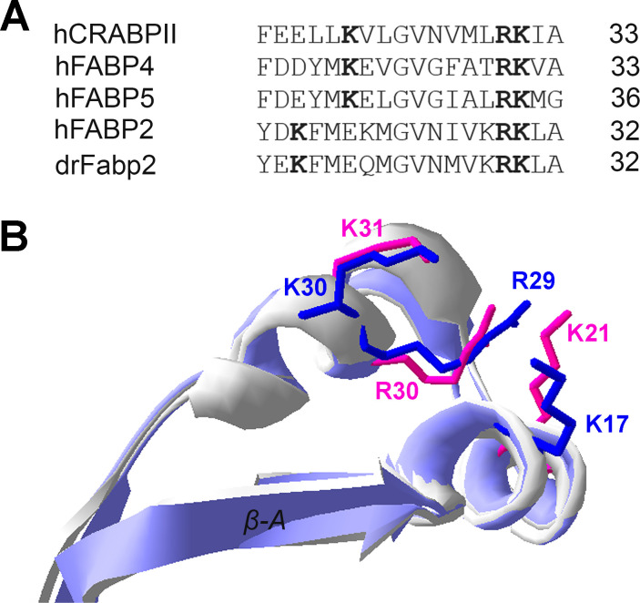
Fig 1 Nuclear localization signal analysis.

A) α-helical regions of ILPs with described NLS (human CRABPII, FABP4 and FABP5) are shown aligned with human (FABP2) and Danio rerio FABP2 (Fabp2). Basic amino acids that compose the NLS are highlighted in bold. B) Superimposed structures of human apoFABP4 (3rzy.pdb) and human apoFABP2 (1kzw.pdb) α-helical region. Side chains of NLS amino acids are shown for human FABP4 (magenta) and human FABP2 (blue).

Acknowledgments
This image is the copyrighted work of the attributed author or publisher, and ZFIN has permission only to display this image to its users. Additional permissions should be obtained from the applicable author or publisher of the image. Full text @ PLoS One