
|
Figure 6
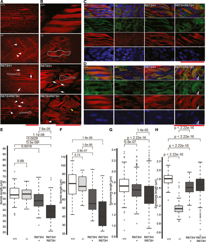
Confocal fluorescence images of slow skeletal muscle of 1 dpf larvae, stained with phalloidin‐rhodamine to detect actin. Puncta of filamentous actin are indicated with white arrowheads, and misshapen myofibers are indicated with white arrows and outlined. Confocal microscope fluorescence images of slow skeletal muscle of 3 dpf larvae, stained with phalloidin‐rhodamine. Puncta of filamentous actin are indicated with white arrowheads, distorted myofibers are indicated with arrows and outlined, and frayed myofibers are indicated with yellow arrowhead. Confocal fluorescence images of slow skeletal muscle of 1 dpf larvae, stained with phalloidin‐rhodamine (red), anti‐α‐actinin antibodies (green), and DAPI (blue). Confocal fluorescence images of slow skeletal muscle of 3 dpf larvae, stained with phalloidin‐rhodamine (red), anti‐α‐actinin antibodies (green), and DAPI (blue). Bundle of actin ringed with α‐actinin indicated with white‐bordered black arrowhead. Myoseptal intervals of slow skeletal muscle at 1 dpf in Myoseptal intervals of slow skeletal muscle at 3 dpf in Z‐disk intervals (sarcomere length) of slow skeletal muscle at 1 dpf in Z‐disk intervals (sarcomere length) of slow skeletal muscle at 3 dpf in
Data Information: Wilcoxon rank‐sum test used to calculate significance.