IMAGE

Fig. 1

ID
ZDB-IMAGE-201003-38
Source
Figures for Nelson et al., 2020
Image
Figure Caption

Fig. 1

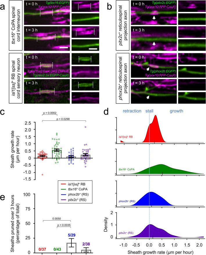
Individual sheath extension rates and pruning frequencies are axon subtype-specific.a-b Lateral view images of the larval spinal cord show sheath extension (a) and sheath pruning (b) during a 3-h time course. Transgenic reporter expression (indicated) marks individual axons or oligodendrocyte membranes. Dashed lines illustrate individual sheath lengths and arrowheads denote sheath pruning events. Images are confocal maximum z-projections with dorsal up and anterior left. Scale bar = 3 μm. c-d Plots show mean growth rates (c) and distributions of individual sheath growth rates (d). For (c), error bars represent the mean ± SEM. For (d), the y-axis approximates the density of data points that exhibited the corresponding growth rate. Larger y-values represent a larger proportion of data points exhibiting this growth rate. Black ticks indicate medians. n (# of sheaths) = 24 (phox2b+), 43 (tbx16+), 36 (pitx2c+), 37 (isl1[ss]+). e Quantification of the percentage of total sheaths that pruned during a 3-h time course grouped by axon subtype. n (sheaths) = 29 (phox2b+), 43 (tbx16+), 38 (pitx2c+), 37 (isl1[ss]+). Overall chi-square test of difference in percent pruning = 0.0025. Pairwise p-values were generated using Dunn’s multiple comparisons (c) or using Tukey-adjusted pairwise comparisons (e)

Acknowledgments
This image is the copyrighted work of the attributed author or publisher, and ZFIN has permission only to display this image to its users. Additional permissions should be obtained from the applicable author or publisher of the image. Full text @ Neural Dev.