
Fig. 7
- ID
- ZDB-IMAGE-200924-2
- Genes
- Antibodies
- Publication
- Zhu et al., 2019 - Zebrafish prmt5 arginine methyltransferase is essential for germ cell development
- All Figures
- Figures for Zhu et al., 2019
|
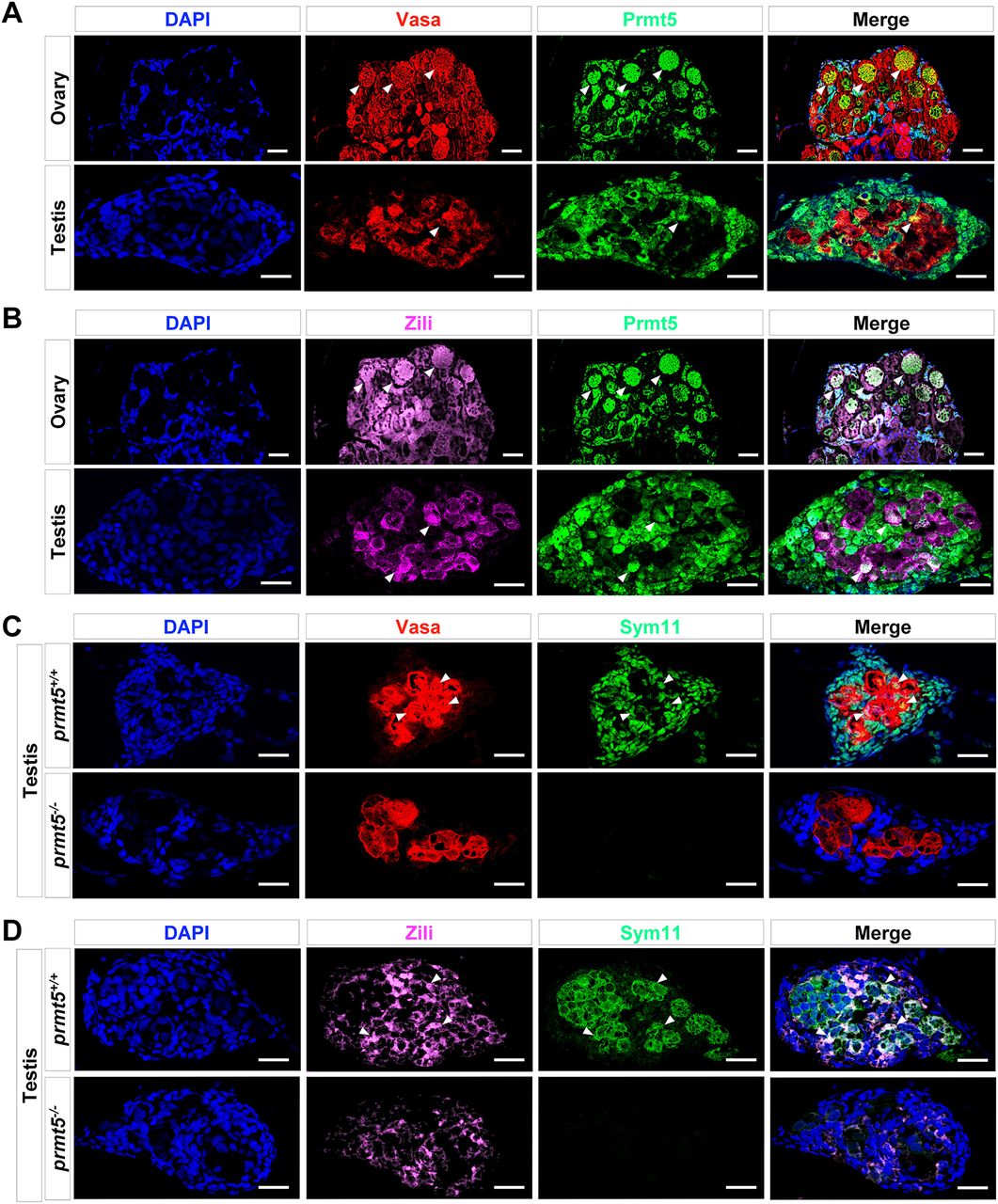
Fig. 7 Co-expression of Prmt5 with Vasa or Zili in both ovaries and testis, and disruption of prmt5 leads to reduction of symmetric dimethylarginine levels in Vasa and Zili. (A) Vasa (red), Prmt5 (green) and DNA (DAPI staining; blue) in wild-type zebrafish ovaries (n=6) and testes (n=6) at 1 mpf. Vasa-positive and Prmt5-positive cells are indicated (white arrowheads). (B) Zili (red), Prmt5 (green) and DNA (DAPI staining; blue) in wild-type zebrafish ovaries (n=6) and testis (n=6) at 1 mpf. Zili-positive and Prmt5-possitive cells are indicated (white arrowheads). (C) Vasa (red), Sym11 (symmetric dimethylarginine antibody, green) and DNA (DAPI staining, blue) in prmt5+/+ testes (n=6) and prmt5−/− gonads (n=6) at 1 mpf. Vasa-positive and Sym11-positive cells are indicated (white arrowheads). (D) Zili (red), Sym11 (green) and DNA (DAPI staining, blue) in prmt5+/+ testes (n=6) and prmt5−/− gonad (n=6) at 1 mpf. Zili-positive and SYM11-positive cells are indicated (white arrowheads). Scale bars: 20 μm.